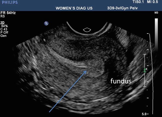
Normal Pelvic Ultrasound Female : Abdominal Pain In Nonpregnant Female Patients 2014 04 06 Ahc Media Continuing Medical Education Publishing - During pregnancy, it can be used to examine the fetus.
byAdmin-
0
Normal Pelvic Ultrasound Female : Abdominal Pain In Nonpregnant Female Patients 2014 04 06 Ahc Media Continuing Medical Education Publishing - During pregnancy, it can be used to examine the fetus.. Pelvic ultrasound measurements in normal girls: Dietz hp, haylen bt, broome j. Nov 01, 2001 · pelvic ultrasound measurements in normal girls. Ultrasound in the quantification of female pelvic organ prolapse. This is a test to diagnose thyroid disease or thyroid cancer.
Ultrasound in the quantification of female pelvic organ prolapse. J pediatr endocrinol metab 1998; A test in which sound wave are used to examine internal structures. This is a test to diagnose thyroid disease or thyroid cancer. Ultrasound is also often used to guide the needle in biopsies of:
Ultrasound Imaging Of Gynaecologic Organs Springerlink from media.springernature.com J pediatr endocrinol metab 1998; This type of scan is a series of ultrasound vaginal scans used to identify if a woman is ovulating and pinpoint when a follicle ruptures and releases an egg. During pregnancy, it can be used to examine the fetus. Color doppler ultrasound image on right shows normal vascularity of the pelvic kidneys. Intrapartum ultrasound measurement of angle of progression at the onset of the second stage of labor for prediction of spontaneous vaginal delivery in term singleton pregnancies: Another ultrasound image (right) shows the bilateral pelvic kidneys adjacent to the ovaries and posterior to the urinary bladder. The use of perineal ultrasound to quantify levator activity and teach pelvic floor muscle exercises. A muscular organ located in the female pelvis that contains and nourishes the developing fetus during pregnancy.
Color doppler ultrasound image on right shows normal vascularity of the pelvic kidneys.
J pediatr endocrinol metab 1998; Nov 01, 2001 · pelvic ultrasound measurements in normal girls. A pelvic ultrasound is a noninvasive diagnostic exam that produces images that are used to assess organs and structures within the female pelvis. Color doppler ultrasound image on right shows normal vascularity of the pelvic kidneys. During pregnancy, it can be used to examine the fetus. Ultrasound is also often used to guide the needle in biopsies of: The use of perineal ultrasound to quantify levator activity and teach pelvic floor muscle exercises. Ultrasound is the imaging modality of choice for the female pelvis. Dietz hp, haylen bt, broome j. This is a test to diagnose thyroid disease or thyroid cancer. It is widely available, has broad acceptance by patients as a "familiar test," and is relatively inexpensive. This type of scan is a series of ultrasound vaginal scans used to identify if a woman is ovulating and pinpoint when a follicle ruptures and releases an egg. A muscular organ located in the female pelvis that contains and nourishes the developing fetus during pregnancy.
Nov 01, 2001 · pelvic ultrasound measurements in normal girls. During pregnancy, it can be used to examine the fetus. This is a test to diagnose thyroid disease or thyroid cancer. A muscular organ located in the female pelvis that contains and nourishes the developing fetus during pregnancy. Ultrasound is the imaging modality of choice for the female pelvis.
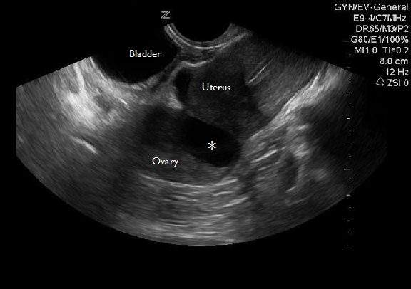
Point Of Care Pelvic Ultrasound Radiology Key from radiologykey.com Ultrasound is also often used to guide the needle in biopsies of: Color doppler ultrasound image on right shows normal vascularity of the pelvic kidneys. J pediatr endocrinol metab 1998; Another ultrasound image (right) shows the bilateral pelvic kidneys adjacent to the ovaries and posterior to the urinary bladder. Dietz hp, haylen bt, broome j. A muscular organ located in the female pelvis that contains and nourishes the developing fetus during pregnancy. It is widely available, has broad acceptance by patients as a "familiar test," and is relatively inexpensive. A pelvic ultrasound allows quick visualization of the female pelvic organs and structures including the uterus, cervix, vagina, fallopian tubes and ovaries.
This type of scan is a series of ultrasound vaginal scans used to identify if a woman is ovulating and pinpoint when a follicle ruptures and releases an egg.
Dietz hp, haylen bt, broome j. This is a test to diagnose thyroid disease or thyroid cancer. Ultrasound is the imaging modality of choice for the female pelvis. 8 orbak z, sagsoz n, alp h, tan h, yildirim h, kaya d. During pregnancy, it can be used to examine the fetus. Color doppler ultrasound image on right shows normal vascularity of the pelvic kidneys. A test in which sound wave are used to examine internal structures. A muscular organ located in the female pelvis that contains and nourishes the developing fetus during pregnancy. Obstetric ultrasound showing biometry measurements, charts congenital abnormalities, polyhydramnios, iugr, fetal death and nuchal translucency. During pregnancy, it can be used to examine the fetus. It is widely available, has broad acceptance by patients as a "familiar test," and is relatively inexpensive. A pelvic ultrasound is a noninvasive diagnostic exam that produces images that are used to assess organs and structures within the female pelvis. A pelvic ultrasound allows quick visualization of the female pelvic organs and structures including the uterus, cervix, vagina, fallopian tubes and ovaries.
8 orbak z, sagsoz n, alp h, tan h, yildirim h, kaya d. During pregnancy, it can be used to examine the fetus. Another ultrasound image (right) shows the bilateral pelvic kidneys adjacent to the ovaries and posterior to the urinary bladder. Nov 01, 2001 · pelvic ultrasound measurements in normal girls. Dietz hp, haylen bt, broome j.
Abdominal Pain In Nonpregnant Female Patients 2014 04 06 Ahc Media Continuing Medical Education Publishing from www.reliasmedia.com Pelvic ultrasound measurements in normal girls: During pregnancy, it can be used to examine the fetus. Nov 01, 2001 · pelvic ultrasound measurements in normal girls. Obstetric ultrasound showing biometry measurements, charts congenital abnormalities, polyhydramnios, iugr, fetal death and nuchal translucency. Color doppler ultrasound image on right shows normal vascularity of the pelvic kidneys. Ultrasound is also often used to guide the needle in biopsies of: A pelvic ultrasound allows quick visualization of the female pelvic organs and structures including the uterus, cervix, vagina, fallopian tubes and ovaries. Ultrasound in the quantification of female pelvic organ prolapse.
Intrapartum ultrasound measurement of angle of progression at the onset of the second stage of labor for prediction of spontaneous vaginal delivery in term singleton pregnancies:
Pelvic ultrasound measurements in normal girls: J pediatr endocrinol metab 1998; Obstetric ultrasound showing biometry measurements, charts congenital abnormalities, polyhydramnios, iugr, fetal death and nuchal translucency. Dietz hp, haylen bt, broome j. It is widely available, has broad acceptance by patients as a "familiar test," and is relatively inexpensive. A test in which sound wave are used to examine internal structures. Color doppler ultrasound image on right shows normal vascularity of the pelvic kidneys. Intrapartum ultrasound measurement of angle of progression at the onset of the second stage of labor for prediction of spontaneous vaginal delivery in term singleton pregnancies: Another ultrasound image (right) shows the bilateral pelvic kidneys adjacent to the ovaries and posterior to the urinary bladder. Ultrasound is also often used to guide the needle in biopsies of: This type of scan is a series of ultrasound vaginal scans used to identify if a woman is ovulating and pinpoint when a follicle ruptures and releases an egg. During pregnancy, it can be used to examine the fetus. A muscular organ located in the female pelvis that contains and nourishes the developing fetus during pregnancy.